No edit summary Tags: Visual edit apiedit |
No edit summary Tags: Visual edit apiedit |
||
| Line 5: | Line 5: | ||
<u>Lungs roots:</u> region of the mediastinum where the lung is connected to the trachea and the heart, consisting of usually a main bronchus, one pulmonary artery and two pulmonary veins. |
<u>Lungs roots:</u> region of the mediastinum where the lung is connected to the trachea and the heart, consisting of usually a main bronchus, one pulmonary artery and two pulmonary veins. |
||
| − | * '''Left root:''' upper part is occupied by left pulmonary artery lying within the cavity of the arch of aorta |
+ | * '''Left root:''' upper part is occupied by left pulmonary artery lying within the cavity of the arch of aorta, arching over the left bronchus. Two pulmonary veins in front and other below the bronchus. |
| − | * '''Right root: '''similar to the left, but bronchus and pulmonary artery branch to the upper lobe divide outside the lung. The artery lies in front of their respective bronchus. |
+ | * '''Right root: '''similar to the left, but bronchus and pulmonary artery branch to the upper lobe divide outside the lung. The artery lies in front and slightly inferior of their respective bronchus. |
<u>Surfaces:</u> the lung generally conform to the shape of the thoracic cavity. |
<u>Surfaces:</u> the lung generally conform to the shape of the thoracic cavity. |
||
Latest revision as of 22:58, 4 September 2015
The lungs are the functional units of respiration, lying within the thoracic cavity. The right lung is composed of three lobes subdivided into 10 segments and the left is composed of two lobes and eight segments. They are surrounded by the pleura which separated them from the chest wall.
The apex of the lung is level with posteromedial first rib. Base is concave adapted to the shape of adjacent diaphragm.
Lungs roots: region of the mediastinum where the lung is connected to the trachea and the heart, consisting of usually a main bronchus, one pulmonary artery and two pulmonary veins.
- Left root: upper part is occupied by left pulmonary artery lying within the cavity of the arch of aorta, arching over the left bronchus. Two pulmonary veins in front and other below the bronchus.
- Right root: similar to the left, but bronchus and pulmonary artery branch to the upper lobe divide outside the lung. The artery lies in front and slightly inferior of their respective bronchus.
Surfaces: the lung generally conform to the shape of the thoracic cavity.
- Costal surface: the interface with the thoracic cage.
- Diaphragmatic surface: the interface with the diaphragm
- Mediastinal surface: On the left, cardiac notch at the anterior margin, arch and ascending aorta make a deep on the lung surface around the hilum, Above the aortic arch are vertical impression made by the subclavian artery and the oesophagus. On the right, cardiac impression is shallower, a groove for the azygos vein over the hilum meets the impression for SCV. Above the arch, is shallow groove for the trachea and right vagus. Inferiorly is the groove for IVC.
- Apical surface: grooved by subclavian arteries.
Borders: three in total.
- Inferior: sharp border that separate the costal and diaphragmatic surface
- Posterior: between medistinal and costal posteriorly, rounded to fit the paravertebral gutters
- Anterior: between mediastinal and costal anteriorly, thin and sharp, and on the left it is deeply concave at the lower part with a cardiac notch.
Fissure:
- Oblique fissure: extends from the anteroinferior surface of the lung to the posterosuperior surface through the hilum dividing the lung into two upper and lower lobes connected only by lobar bronchi and vessels.
- Horizontal fissure: passes from the anterior margin into the oblique fissure to separate a wedge shaped middle lobe from the upper lobe.
Lobes:
- Bifurcation of the trachea into each main bronchus passes downwards and laterally to enter the hilum of the lung. The right main bronchus is shorter wider and more vertical than the left.
- each bronchus becomes lobar bronchi that supply the lobes of the lung.
- right bronchus gives upper lobe bronchus outside the hilum, and divide into middle and lower lobe bronchi within the hilum. Left main bronchus divides within the hilum to upper and lower lobar bronchi.
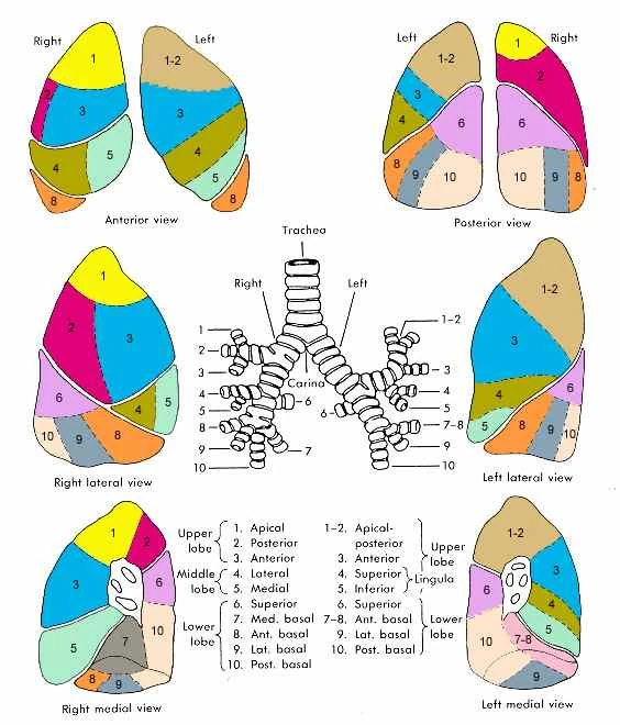
Segments:
- right lung: upper lobe is apical, posterior and anterior. Middle lobe is medial and lateral. Lower lobe is superior, medial, basal, anterior basal, lateral basal and posterior basal.
- left lung: upper lobe is apical, posterior, anterior. Lower lobe is superior lingular and inferior lingular, and superior, medial basal, anterior basal, lateral basal, posterior basal.
- superior segment of the lower lobe is supplied by a bronchus which is highest to arise from the posterior surface of the bronchial tree, so material aspirated by a supine patient would tend to gravitate into this segment.
Blood supply
The lungs have dual arterial supply and venous drainage: pulmonary arteries and veins as well as bronchial arteries and veins:
- arterial supply
- pulmonary arteries: supply de-oxygenated blood from the right ventricle. Divides with the bronchus but does not supply it, supply the alveoli instead.
- bronchial arteries: two branches of the thoracic aorta (on left) and one from the third right posterior intercostal artery (on right) that supply oxygenated blood to the bronchial tree from carina to respiratory bronchioles, and visceral pleura.
- venous drainage
- pulmonary veins: drains to the left atrium. These are formed from tributaries but do not follow the bronchi, rather follow in the intersegmental septa.
- bronchial veins: superficial system drains into azygos and systems while deep system drain into main pulmonary vein.
Nerve supply
- Pulmonary plexuses situated anterior and posterior to the hilar structures receive autonomic nerve from cardiac plexuses and upper four thoracic sympathetic ganglian and thoracic vagus.
- Vagus (parasympathetic ) fibres are afferent (for cough reflex) and efferent (bronchoconstrictor, vasodilator, secretomotor to glands). sympathetic efferents are bronchodilators and vasoconstrictors.
Lymphatic supply
The lymphatics drain via superficial and deep lymphatic plexuses to bronchopulmonary nodes at the hilum. The superficial lymphatic plexus is subpleural while the deep lymphatic plexus accompanies the bronchovascular structures with associated intrapulmonary lymph nodes.
Variant anatomy
- bronchopulmonary foregut malformations
- accessory lobes - may not have own segmental supply, e.g. accessory inferior lobe
- cardiac bronchus - from right bronchus
- supernumerary tracheal bronchus - segmental bronchus arises from the trachea
- oesophageal lung (extremely rare)
- main bronchus arises from the oesophagus
- pulmonary vascular supply
- pulmonary aplasia
- ectopic lung tissue - most common trachea
- agenesis of unilateral lung
- Fissure variants:
- azygos fissure: most commonly seen accessory fissure
- Normal variant seen in approximately 0.5% of chest radiographs
- Reported 2:1 male to female ratio
- inferior accessory fissure
- superior accessory fissure
- left horizontal fissure
- azygos fissure: most commonly seen accessory fissure